AI-Powered
Brain Tumor Detection
Empowering neurologists and medical professionals with real-time, high-accuracy MRI analysis using specialized Deep Learning models and advanced segmentation.
0+
Diagnostic MRI Scans Processed
0.0%
Detection Accuracy
< 0.0s
Real-Time Analysis
256-bit
Secure Encrypted Storage
Platform Capabilities
A comprehensive suite of AI tools designed to integrate seamlessly into modern neuro-diagnostic workflows.
AI Tumor Detection
Powered by YOLOv11s-seg, our model identifies and classifies gliomas, meningiomas, and pituitary tumors instantly — the core engine behind every scan on this platform.
Smart MRI Upload
Securely upload DICOM, JPG, or PNG scans. Our preprocessing pipeline automatically normalizes images for optimal AI analysis.
Precise Segmentation
Go beyond detection. The AI draws pixel-perfect bounding boxes and segmented masks to isolate tumor mass boundaries.
Validation Guardrail
An integrated MobileNetV3 model acts as a security layer, ensuring only valid brain MRI scans are processed by the core engine.
AI Medical Chatbot
Interact with our DistilBERT-powered assistant for instant querying of platform knowledge and medical terminology support.
Clinical PDF Reports
Generate and download comprehensive, timestamped diagnostic reports ready for patient files or secondary doctor review.
Seamless Clinical Workflow
From upload to diagnosis in four simple steps.
Upload Scan
Upload a patient's brain MRI via our secure, encrypted dashboard interface.
Guardrail Check
System validates the image to ensure it is a legitimate anatomical MRI scan.
AI Processing
Deep learning models analyze the scan, extracting anomalies and segmenting tumors.
Review & Export
Doctor reviews the visual output, adds clinical notes, and downloads the official PDF.
Research-Grade Precision
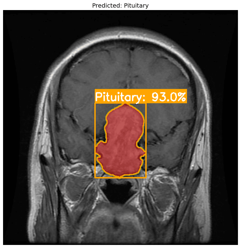
Visualizing the raw power of our customized YOLOv11 instance segmentation engine in isolating complex brain anomalies.
Multi-Class Identification
Accurately distinguishes spatial variations between Gliomas, Meningiomas, and Pituitary tumors.
Pixel-Level Instance Masking
Generates exact irregular coordinates of tumor mass volume borders rather than simple rigid box outlines.
Confidence Layer Matrix
Provides clear statistical probability scores directly integrated alongside diagnostic bounding coordinates.
24/7
Automated availability for emergency radiologist support.
0+
Major tumor classes identified simultaneously.
Why Choose MedVision AI?
We bridge the gap between cutting-edge artificial intelligence and practical clinical application. Our platform acts as a powerful second opinion tool, reducing diagnostic fatigue and accelerating patient care timelines.
- Accelerate diagnosis from days to milliseconds.
- HIPAA-compliant architecture and data anonymization.
- Maintain persistent, searchable patient diagnostic histories.
Trusted by Professionals
Hear from the researchers and clinicians using MedVision AI.
MedVision acts as an incredible preliminary screening tool. The segmentation accuracy on low-contrast gliomas is highly impressive.
Dr. Sarah J.
Neurologist
The integration of a safeguard model to reject invalid imagery saves our research team countless hours of data cleaning.
Marcus T.
AI Research Lead
Generating automated PDF reports directly from the inference engine completely streamlines our daily diagnostic workflow.
Dr. Ahmed R.
Chief Radiologist
Frequently Asked Questions
Absolutely not. MedVision AI is designed as a clinical decision support system (CDSS). It acts as a highly accurate 'second opinion' to assist radiologists and reduce diagnostic oversight.
Currently, the platform accepts standard image formats like JPG and PNG for the demo. Enterprise deployments support native DICOM medical formats.
Our customized YOLOv11s-seg model has been trained on thousands of varied MRI scans, achieving over 96% accuracy in detecting and segmenting core tumor classes.
Yes. Once an analysis is complete, doctors can add clinical notes and export a timestamped, professional PDF report instantly.
Security is our top priority. We use robust encryption and do not share your medical scans with third-party advertisers.